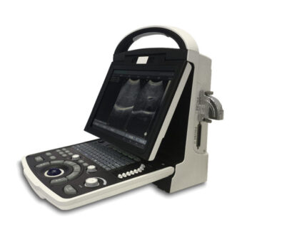
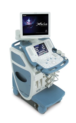
Toshiba Xario Prime Ultrasound
Toshiba Xario Prime Ultrasound Machines brings premium technology to an affordable platform of compact, stylish design. Xario combines the highest level of performance with ergonomic design to meet the fast-paced demands of your daily practice with speed and diagnostic confidence.
Click here to download the product catalog
Toshiba_Xario.pdf
PLEASE CLICK HERE TO SEE OUR FULL RANGE OF ULTRASOUND MACHINES
Toshiba Xario Prime Ultrasound has powerful system architecture, whose intelligent components work and communicate autonomously, supports the most advanced imaging functions. Xario is also easy to upgrade, to keep you abreast of new ultrasound techniques.
Excellent diagnostic performance. Operator comfort that inspires productivity. And outstanding connectivity features. Xario. Quite simply, the prime ultrasound system for a wide range of clinical applications.
Toshiba Xario Prime Ultrasound offers unsurpassed image quality, backed by unique, clinically proven technologies. Its full range of advanced imaging functions lets you visualize minute tissue details and vascular structures with precision, for a faster, more accurate diagnosis. Xario's transducers deliver superb image quality and respond with outstanding versatility to a wider range of applications
XBT Transducer Technology
Transducers of unsurpassed bandwidth and sensitivity
enable ultrasound images of both high spatial resolution and increased penetration. One transducer can be used in a wider range of applications for greater cost efficiency.
Pulse Subtraction THI
Optimizes spatial and contrast resolution in grayscale
imaging. A standard feature with all Xario transducers.
ApliPure
Real-time compounding delivers images of outstanding
clarity and detail, while preserving clinically significant markers.
Advanced DYNAMIC FLOW
Brings superior spatial resolution to Color Doppler.
Depicts even tiny vessels and flow around plaques with improved accuracy and detail.
Low MI Pulse Subtraction
Real-time Contrast Imaging with outstanding sensitivity
in an easy-to-use package for daily practice.
Trapezoid Imaging
Extends the field of view for a better overview of the region
of interest in both grayscale and Color Doppler modes.
Panoramic View
Reconstructs a single wide-view frame from continuous
ultrasonic images for improved visualization of widespread regions and anatomical relationships.
Fast Fusion 3D
Provides clear 3D images of complex structures, such as
tumors and their feeding vessels, with simple operation.