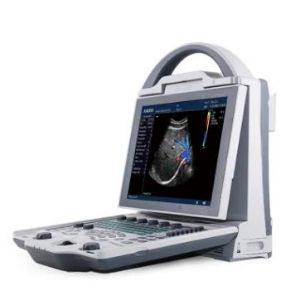
Ultrasound Machine DCU12 Color Doppler
Ultrasound Machine DCU12 Color Doppler is a portable color doppler, designed and produced by
KAIXIN Medical. The Leading Image process technical and advanced plus practical clinic Solution Method. It's highly cost effective and reliable machine. Kaixin Ultrasound Machine DCU12 Color Doppler is ideal for hospitals and clinics. For more information regarding the machine please feel free to contact us at +923224602203 or Email: info@zirarenterprises.com, zirarenterprises@gmail.com.
DOWNLOAD COMPLETE CATALOG HERE KAIXIN DCU12 Brochure.pdf
Whats in the Box:
- 1 x Kaixin Ultrasound Machine
- 1 x Convex Probe
- 1 x Linear Probe
- 1 x Standard Accessories
- 1 x Trolley (Free)
Optional Configuration:
- Tvs Probe @150,000 (optional)
https://www.youtube.com/watch?v=cf3KqfxAIow
1. Application:
Leading image process technical and advanced and practical clinical solution method. Applicable to diagnosis of abdominal organs(trans-vaginal included), superficial organs and cardiac. With scale-able design, upgrade continuously, fully meet the needs of clinical application extension.
2. Parameters:
Specification parameters 1. Grey scale: 256 level
2. Color scale: 256 level
3. Display: 10.4'flicker-free high-resolution medical color LCD
4. Power: 100-240V, 1.2-0.6A frequency: 50-60Hz
5. Adapter output: DC12.8V 3.0A
6. Power consumption: ≤100VA
7. Main unit size: 256*150*326(mm, Length*width*height)
8. Unit net weight: 4.5Kg(Accessories not included)
Multi-media and accessory equipments 1. Video recorder
2. I-station integral work station, realize image storage, report form and cloud print
3. U flash disk (file management, software upgrade and one-key storage) and DICOM port, convenient for data management and available to removal transmission.
4. Dual-mode TV output: PAL/NTSC
Probe port Two enabled probe ports, automatic identification technology, available to optional probe configuration.
Language Menu operation, interface language: English/Chinese switch alternatively
3. Operation Mode:
B,B/B,4B
M,B/M
CFM
PDI
PW
THI
4. Ultrasonic imaging technology:
- High precise digital multi-beam formator
- Dynamic frequency fusion imaging
- High precise delay dynamic receiving focusing
- Ultra-wideband image technique
- Adaptive color artifact removal technique
- Adaptive vessel imaging
- Adaptive Doppler image technique
- THL tissue harmonic imaging technology
5. Measurement report:
OB measurement report, GYN measurement report, cardiac measurement report, urology measurement report and other measurement report, automatically store the measurement result and generate report.
6. Configuration:
6.1 Standard Configuration:
1 Main unit:1 pc
2 3.5 MHz convex abdomen probe:1 pc
3 You can choose 1pc probe from below:
>6.5 MHz trans-vaginal probe
> 7.5 MHz linear probe
4 Workstation software: 1 set
5 Reticle:1 pc
6.2 Optional Cofiguration
1 Foot Switch
2 Trolley
3 Video Recorder